What is Glaucoma?
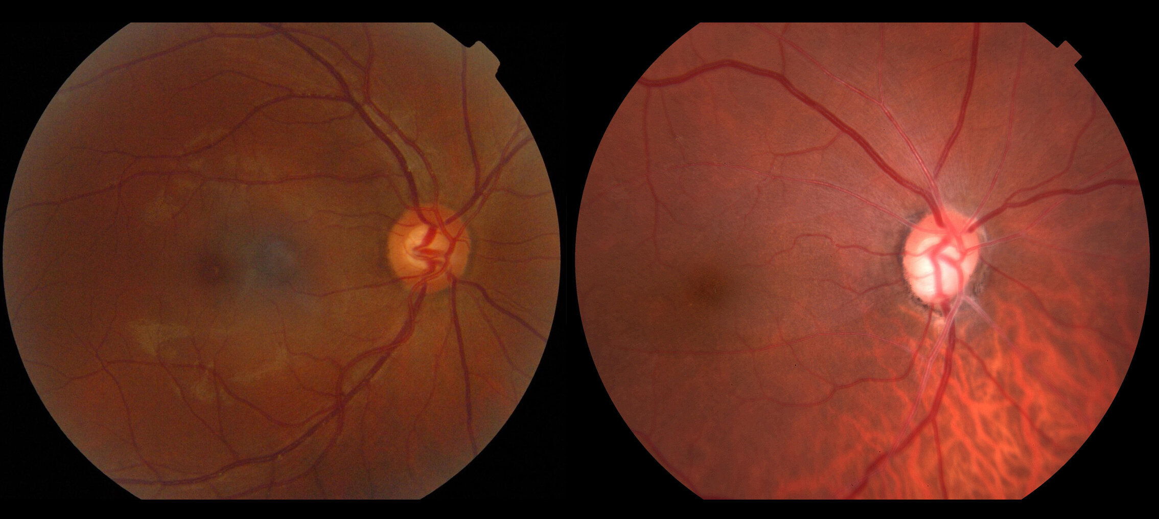
Fig. Normal optic nerve (Left) compared to a glaucomatous optic nerve (Right).
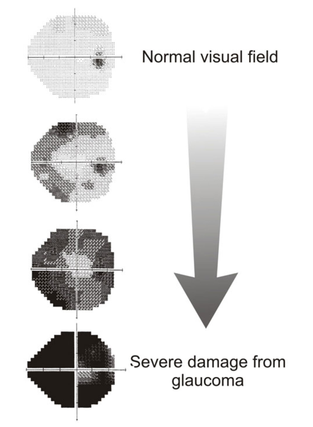
Glaucoma encompasses a group of eye conditions which cause damage to the optic nerve, the structure that carries nerve fibres from the eye to the brain. This results in irreversible vision loss starting in the periphery then slowly involving your central vision. being lost irreversibly. The main risk factor for the development and progression of glaucoma is elevated pressure within the eye.
What causes pressure within the eye?
Eye pressure is controlled by aqueous humour, fluid which fills the front part of the eye and drains through tiny channels, called the trabecular meshwork.
In a normal eye, there is a balance between the production and drainage of this fluid, but in glaucoma this balance is disturbed, which typically causes the eye pressure to rise. This is usually because the drainage of fluid from the eye becomes restricted.
Normal eye pressures ranges from 10-21 mm Hg. However, there is no specific level of elevated eye pressure that definitely leads to glaucoma; conversely, there is no lower level of pressure that will absolutely eliminate a person’s risk of developing glaucoma. Every optic nerve is unique and has a different eye pressure that it can tolerate. That is why early diagnosis and individualised treatment of glaucoma is the key to preventing vision loss.